CRYSTAL VISION OPTOMETRY
  • Home
  • Our Practice
    • Contact & Location
    • Testimonials
  • Our Services
    • Orthokeratology Myopia Control >
      • Orthokeratology Infromation
    • Comprehensive Optometric Eyecare
    • Contact Lens Evaluation
    • Specialty Contact Lenses/Scleral
    • HOA Scleral Contact Lenses
    • IPL For Dry Eye
    • IPL For Chalazion
    • Red Eye-Ocular Emergencies
    • Glaucoma Treatments
    • Lasik Consultation and Post OP Care
    • Pediatric Eye Care
  • Patient Center
    • Patient Forms
    • Insurance and Payments
    • Promotions
    • Informational Videos
    • Appointment Scheduling
  • Innovative Technology
    • Medmont Topographer
    • Pentacam Tomographer
    • Optikam
    • Zeiss OCT
    • Eidon Retinal Scanner
  • Eye Care Articles

Pentacam  Tomographer

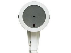
                               PENTACAM  TOmOGRAPHER

Picture
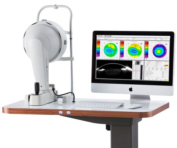
​​Here at Crystal Vision Optometry in Fremont, CA We have the latest innovative Technologies in all our equipment such as The Pentacam, Corneal tomography and Scheimpflug imaging.
​
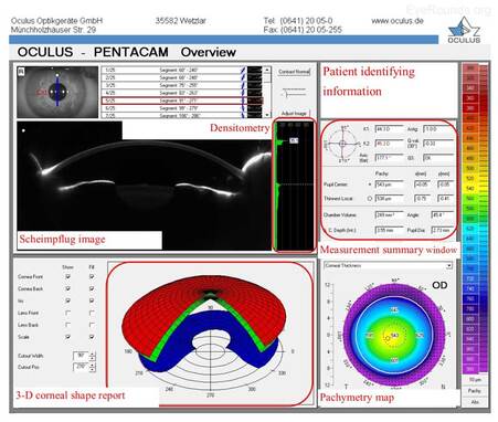
It is frequently used to analyze the corneal surface, especially in the field of cataract and refractive surgery. The Pentacam is one of the most commonly used commercially available systems for this purpose. Through a rotating Scheimpflug camera, the system is capable of creating a three-dimensional map of the cornea. These advances in tomography have simultaneously enhanced the ability of clinicians to screen surgical candidates and detect subtle corneal changes in diseases such as keratoconus. 
However, there remains a need to enhance diagnosis in order to recognize mild and early forms of corneal ectasia. As iatrogenic ectasia and keratoconus are dreaded complications of refractive surgery, it is imperative to screen patients appropriately prior to surgery.

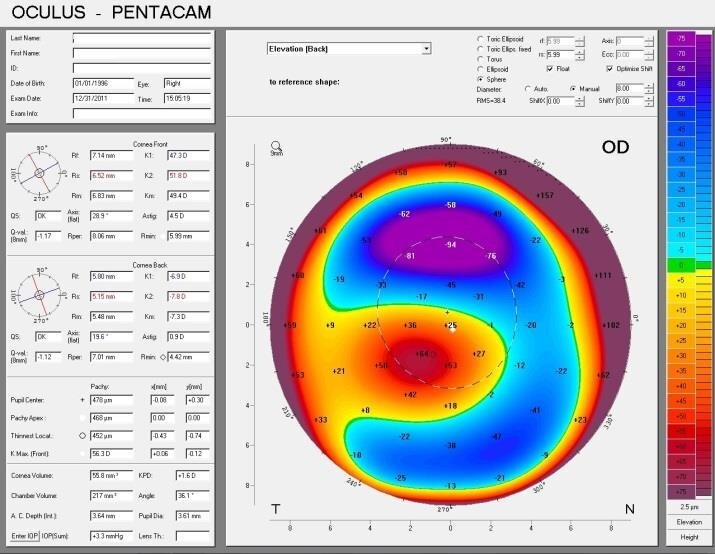
The Pentacam is one of many systems utilized in the screening process, but the literature has not identified specific protocol nor parameters that are capable of carrying out this process appropriately. Post-operative keratoconus continues to occur despite the advances in technology seen in corneal imaging. Therefore, clear indices for screening are required in order to diagnose early forms of keratoconus and other corneal diseases that may exclude the seemingly asymptomatic patient from undergoing refractive surgery. This article aims to summarize the indices available on the Pentacam system and to identify the most accurate parameters for screening of the refractive surgery candidate.

Picture
Picture
Picture
Clinical uses of Pentacam
  • Screening for corneal ectasiaKeratoconus, the most common corneal ectasia, is a progressive corneal condition characterized by central thinning and steepening of the cornea. Early keratoconus often looks normal on slit lamp examination, and manual keratometry, which assesses the central 3 mm, may give an insufficient assessment. Because of this, topography has become the gold standard for screening patients for keratoconus and other corneal ectasias (Figures 6-8).
  • Corneal ectasia monitoring and treatment,Once an ectasia (e.g., keratoconus, Pellucid marginal corneal degeneration) is diagnosed, topography may be useful for monitoring disease progression. With regular surveillance topography, it can be determined when patients are at risk for progression and complications, and this precise monitoring allows early intervention with treatments such as collagen cross-linking or keratoplasty. Topographic warning signs include high central corneal power, a large difference between the two corneas of a patient, and a large disparity between the refractive power at the apex and the periphery (Figures 6-8) (5).
  • Refractive surgery screening and monitoring, Laser refractive surgeries such as photorefractive keratectomy (PRK) and laser assisted in situ keratomileusis (LASIK) use excimer laser to ablate tissue and reshape the cornea to correct an individual's refractive error.  Not every patient, however, can safely undergo these procedures. Screening must be performed to determine corneal shape and patterns of astigmatism on topography before refractive surgery can safely be performed. Topography can also be used post-operatively to evaluate etiology for unsatisfactory visual outcome such as decentered or incomplete ablations.
  • Pre-operative intraocular lens selectionDuring cataract surgery, an intraocular lens is placed in the eye to achieve the desired refractive outcome.  Standard intraocular lenses only contain spherical correction.  If a patient has regular corneal astigmatism, however, an astigmatism-correcting toric lens may be used.  Corneal topography is a useful pre-operative test to assess the magnitude and regularity of corneal cylinder when selecting an intraocular lens implant prior to cataract surgery.
  • Post-keratoplasty astigmatism evaluation and managementAfter keratoplasty, corneal astigmatism can be evaluated with topography. This technology guides selective suture removal and other interventions to reduce levels of astigmatism.
  • Ocular surface disorder evaluation, Ocular surface disorders, such as pterygia, corneal scars, and Salzmann nodules, can induce irregular corneal astigmatism. Corneal topography can be used to evaluate the refractive effects of these problems and to aid in disease monitoring and surgical planning.

Book Your Appointment
Contact Us
5395 Mowry Ave. Fremont, CA 94538
Phone: 510-440-9825 
Fax:  
510-250-1065
​Email: [email protected]
Temporary Office Hours
Until Further Notice
Mon, Tues, Thurs, Fri**  9:30 am - 6:00 pm

Wed  Closed
Every  Saturday**   9:00 am - 1:00pm
**By appointment only
**Can only book by call not online


Picture
Powered By GotWebCo.com
  • Home
  • Our Practice
    • Contact & Location
    • Testimonials
  • Our Services
    • Orthokeratology Myopia Control >
      • Orthokeratology Infromation
    • Comprehensive Optometric Eyecare
    • Contact Lens Evaluation
    • Specialty Contact Lenses/Scleral
    • HOA Scleral Contact Lenses
    • IPL For Dry Eye
    • IPL For Chalazion
    • Red Eye-Ocular Emergencies
    • Glaucoma Treatments
    • Lasik Consultation and Post OP Care
    • Pediatric Eye Care
  • Patient Center
    • Patient Forms
    • Insurance and Payments
    • Promotions
    • Informational Videos
    • Appointment Scheduling
  • Innovative Technology
    • Medmont Topographer
    • Pentacam Tomographer
    • Optikam
    • Zeiss OCT
    • Eidon Retinal Scanner
  • Eye Care Articles