zeiss oct
|
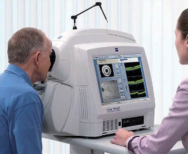
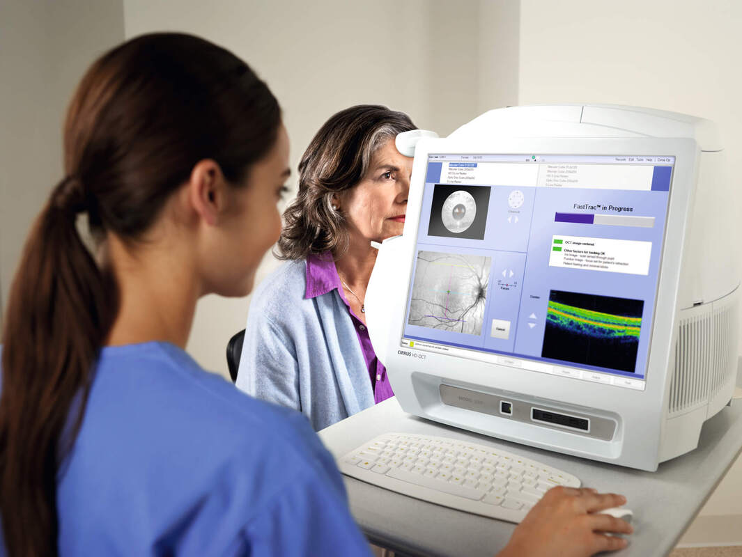
Over 25 years ago, Optical Coherence Tomography (OCT) revolutionized eye care by allowing doctors to see into the eye like never before. Now OCT is routinely used here at Crystal Vision Optometry for eye exams and is a standard of care in ophthalmology and optometry. A quick non-invasive OCT exam reveals details which help doctors detect potentially debilitating eye diseases, like macular degeneration, glaucoma or diabetic retinopathy, and supports them in making treatment decisions. Doctors have found that early diagnosis and treatment may help to preserve vision.
OCT had its origins in academic research and was patented at Massachusetts Institute of Technology (MIT) in 1991. |
In collaboration with MIT’s leading researchers, clinical scientists and engineering experts, ZEISS played a key role in bringing this groundbreaking technology to eye care by building the first commercially available OCT device in 1996. Today OCT technology continues to advance eye care with new applications and technology innovations that provide even more avenues for better diagnosis and treatment of ophthalmic diseases.
|
But what exactly is OCT and how does it work?
Early detection of serious eye conditions, such as retinal detachment or glaucoma, can significantly decrease a patient’s risk of vision loss. Optical Coherence Tomography (OCT) is an imaging technology that uses light waves to capture detailed, cross-sectional sub-surface images of transparent material. This imaging technology is used throughout the medical world in oncology, cardiology, dermatology, neurology and, most widely, in ophthalmology. OCT is a breakthrough in eye care that has allowed doctors to more easily detect eye disorders. In ophthalmology, OCT is used to capture detailed images of the eye. The information from OCT images helps ophthalmologists and optometrists to diagnose and manage potentially damaging ocular diseases which, if left untreated, can lead to blindness or seriously impact a person’s quality of life. OCT functions much like an ultrasound system, but has much higher resolution. Where ultrasound systems use sound waves to capture images, OCT uses infrared light that is similar to the light of a TV remote control device. In eye care, OCT imaging uses the light reflected from the retina to create extremely high-definition volumetric 3D scans that detail various layers of the eye. Thanks to the use of light, OCT systems can capture details thinner than a human hair, helping doctors detect abnormalities which may otherwise remain unseen. These two or three-dimensional OCT scans produce brilliant, high-resolution digital images of the eye and capture much more than standard photographic imaging ever could. Along with this visual information, OCT systems also provide analytics giving clinicians insights into evolving structural changes in the eye. Doctors can analyze this information to more easily diagnose eye diseases earlier and better discern changes associated with disease than was possible before the advent of OCT. OCT provided an entirely new way to look into the eye, making it possible to see details inside the structure of the eye which were previously not visible. Many patients may not notice symptoms of eye disease. OCT helps doctors identify anomalies in ocular structures and detect diseases that may threaten sight. OCT can also aid doctors in determining the best treatment option for their patient and can help to track the progression of eye disease. OCT has been, and is continuing, to be a rapidly evolving field helping to advance eye care. A recent major advance in OCT is OCT Angiography which allows imaging of the vascular structures of the eye, reducing the need for more invasive imaging approaches, such as fluorescein angiography. OCT Angiography provides non-invasive 3D images of blood flow in the eye to help clinicians detect retinal diseases earlier, such as diabetic retinopathy and macular degeneration. Swept-Source OCT (SS-OCT) is another emerging technology, which uses a short cavity swept laser with longer wavelengths and a high-speed camera, to give clinical researchers the potential to see deeper, wider, and in more detail, aiding better assessment of a patient’s treatment response and monitoring of diseases, such as diabetic retinopathy and wet age-related macular degeneration (AMD). Within ophthalmology, another major expansion has been into surgical microscopes enabling 3D visualization of the retinal structures during surgery, helping to potentially enable more accurate surgery and potentially reducing trauma to the eye. OCT technology is not just for eye care, researchers are investigating the use of OCT in neurological diseases, such as Multiple Sclerosis, Parkinson’s and Alzheimer’s. OCT is also used in other medical applications such as neurology, cardiology, oncology, and dermatology. |
